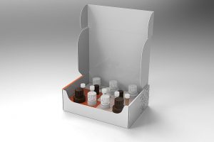
| Detection method | Colorimetric (OD570-573nm) |
| Assay type | Quantitative |
| Samples type | Tissues / Cells |
| Species | All |
| Guideline for sample volume | 20 mg of tissue per 1 mL of extraction buffer 0.5 M cells per 100 μL of extraction buffer |
| Metabolites measured | NAD+, NADH |
| Detection Limit | 25 nM for NAD+ and 35 nM NADH in the cell/tissue extract |
| Assay Length | < 1h |
| Size | 192 reactions |
| Number of samples | 40 (measured in duplicates) |
| Storage | Store reagents at -80°C |
| Intended use | RUO |
| Shelf Life | 6 months |
Key Features
- Highly accurate and sensitive.
- Easy to handle. One step metabolite extraction is followed by separate measurement of NAPD+ and NADPH in the extract. Detection time is less than 10 minutes.
- High throughput. Can be easily adopted into high-throughput 96-well plate assay to analyze hundreds of samples per day.
Principals
The principle of the assay is a cyclic enzymatic reaction with a colorimetric end-point detection. First, NADP+ and NADPH metabolites are extracted together from a tissue or cell culture sample in a single step. Then the extract is divided into two parts revealing both NADP+ and NADPH concentrations by removing one metabolite and stabilizing the other.
Reagents Provided
Extraction BUFFER A, NAPD+ stabilizing reagent, NADPH stabilizing reagent, NADP+ standard stock, NADPH standard stock, Assay BUFFER C, Assay color reagent, Enzyme, Stop solution, Positive control
Kit Requires
Multi-channel pipets. Two clear-bottom 96-well plates suitable for colorimetric assays, heat block with adjustable temperature (up to 80°C), spectrophotometric plate reader
Precautions and Warnings
The Stop Solution may cause skin, eye, and respiratory irritation. Avoid breathing in fumes.
Assay color reagent may cause skin irritation. Handle with care, use gloves.
BUFFER A can cause eye irritation. Handle with care, use googles.
Read more in Q-NAD Safety Data Sheet (SDS), last modified January 2024
General Information
Proprietary name: Q-NADP tissue/cells: quantitative assay kit for NADP+ and NADPH in tissues and cells
More details how to prepare samples in sample preparation document.

